Engineers are available to assist.

Optical components from Edmund Optics® are used in countless Life Science-related applications and medical devices such as qPCR instruments, fluorescence microscopes (confocal, multi-photon, super-resolution), flow cytometers (cell sorting), ophthalmoscopes, optical coherence tomography (OCT), and many more. The optics in such devices enable the detection, diagnosis and treatment of conditions such as COVID-19, cancers, macular degeneration, diabetic retinopathy, glaucoma, genetic disorders, hormonal imbalances in the brain, apoptosis in the blood, etc.
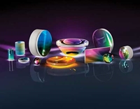
Edmund Optics offers all of the optical components you need for building medical devices such as high precision filters, lenses, mirrors, beamsplitters, polarizers, microscope objectives, and more. In addition, Edmund Optics' portfolio comprises a range of light sources and cameras to complete your setup whether you are designing a microscope, an OCT system, or a different type of optical medical device.
Flow Cytometry is an analytical technique used in a variety of life science applications for counting, inspecting or sorting particles in solution such as single cells. This technique enables the analysis of mixed cell populations - for example from blood, bone marrow, or even from solid tissues such as tumours when these are dissociated into single cells. Flow cytometry is used in a variety of disciplines such as immunology, cancer, virology and molecular biology, as well as infectious disease monitoring. Flow cytometers consist of lenses, optical filters, mirrors, prisms and other optical components to direct the light which are crucial in the successful operation of these systems.
Learn More
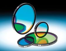
Optical Filters
VIEW ALL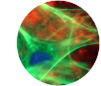
Fluorescence imaging is a powerful, highly sensitive and non-invasive technique used in life sciences to visualise and monitor biological processes in live or fixed cells, tissues, or even complete organisms. Fluorescence is the emission of radiation, visible or invisible, by a substance such as a fluorescent dye (also called fluorophores or chromophores), upon excitation with light or other electromagnetic radiation. There are a wide range of fluorescent dyes and proteins commercially available that can be used to label biological structures with high specificity. Fluorescence-based techniques are varied and include qPCR, DNA sequencing and many microscopy techniques such as confocal, multiphoton and light sheet microscopy. Fluorescence filter sets are particularly important in separating the excitation light from the emitted fluorescence and commonly consist of a combination of an excitation, dichroic and emission filter with transmission profiles optimised to match the spectral characteristics of certain fluorophores.
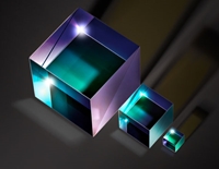
Learn MoreOptical coherence tomography (OCT) is a non-invasive, high-resolution optical imaging technology that creates cross-sectional images from interference signals received from an object under investigation and a reference optic. OCT allows improved diagnosis of ophthalmic diseases, such as Age-related Macular Degeneration, AMD (a disease of the retina), which causes blurred vision or diabetic retinopathy, by quantitatively characterising changes in the structure and appearance of retinal tissue. Edmund Optics supplies a wide range of optics ideal for OCT systems, including plate and cube beamsplitters, broadband dielectric mirrors, lenses, illumination sources and whole Lumedica benchtop OCT Imaging Systems.
Learn MoreThe manipulation of light is critical in the fight against COVID-19, which is caused by the novel coronavirus. Frontline healthcare workers and researchers use systems composed of a wide variety of optical components including filters, mirrors, lenses, objectives, and more. These systems include polymerase chain reaction (PCR) devices that replicate DNA samples for testing, antibody detection systems, infrared fever detection devices, UV cleaning robots, and systems for monitoring the blood of patients connected to ventilators.
Learn More


or view regional numbers
QUOTE TOOL
enter stock numbers to begin
Copyright 2023, Edmund Optics Inc., 18 Woodlands Loop #04-00, Singapore 738100
California Consumer Privacy Act (CCPA): Do Not Sell or Share My Personal Information
California Transparency in Supply Chains Act