Engineers are available to assist.


Fluorescence imaging is a powerful, highly sensitive and non-invasive technique used in life sciences to visualize and monitor biological processes in live or fixed cells, tissues, or even complete organisms. Fluorescence is the emission of radiation, visible or invisible, by a substance such as a fluorescent dye (also called fluorophores or chromophores), upon excitation with light or other electromagnetic radiation. There are a wide range of fluorescent dyes and proteins commercially available that can be used to label biological structures with high specificity. Fluorescence imaging techniques can be divided into spectroscopic and microscopic fluorescence methods. Applications based on spectral detection include qPCR, DNA sequencing, high-throughput screening, or flow cytometry. Fluorescence microscopy techniques commonly used in biomedical research laboratories include epifluorescence, confocal, multiphoton, TIRF (total internal reflectance fluorescence), super-resolution (SIM, STORM, PALM, STED), or light sheet microscopy, as well as fluorescence lifetime imaging. The most suitable technique will depend on multiple factors including the level of two- or three-dimensional resolution, imaging speed, imaging depth, or the number of colour channels required, but all utilize the same fluorescence mechanism to observe biological processes.
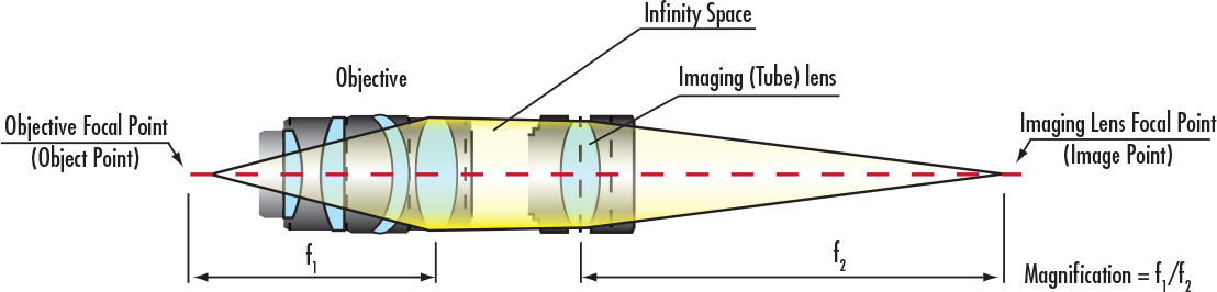
A typical fluorescence imaging system consists of the following components:
Fluorescence filter sets are particularly important in separating the excitation light from the emitted fluorescence and commonly consist of a combination of an excitation (typically bandpass), dichroic (sometimes referred to as dichroic mirror or dichromatic beamsplitter), and emission (bandpass or longpass) filter with transmission profiles optimized to match the spectral characteristics of certain fluorophores.
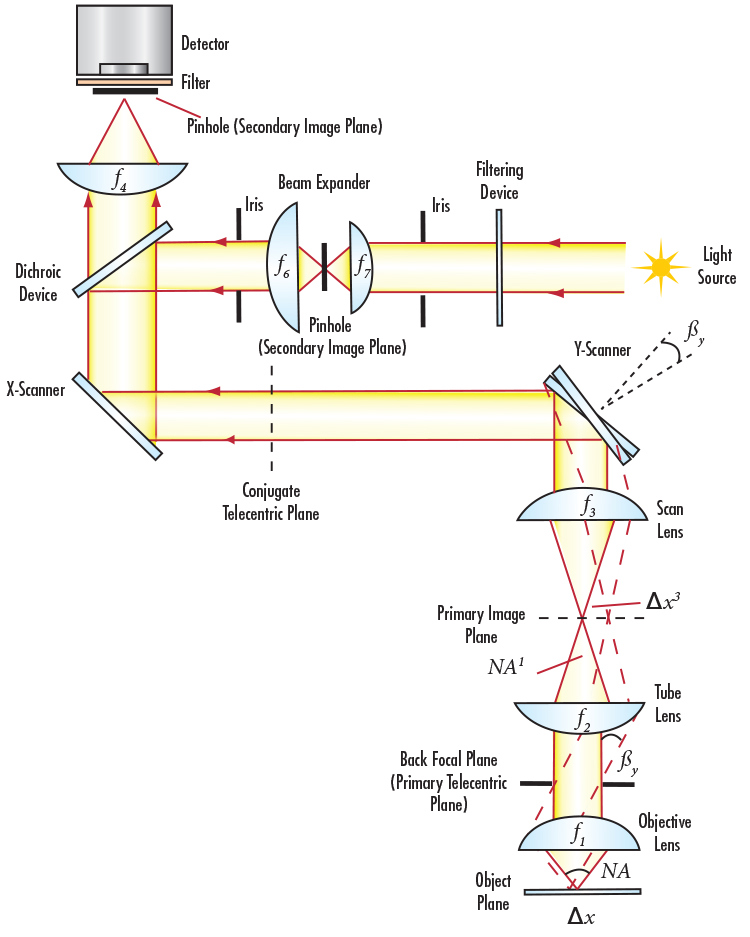
Fluorescence microscopy is one of the primary techniques used to study the functional or morphological dynamics of synaptic structures, including dendritic spines and axon terminals, and to characterize circuit connectivity and dynamics. Typically, a laser beam is focused into a pinhole, acting as a point source illuminator. Spatially filtered light is reflected by a dichroic filter, which may or may not require a beam expander to fill the entire aperture of the objective. The objective then focuses excitation energy onto the sample, which emits a weaker fluorescence signal collected by the same objective. This emission light is transmitted through the dichroic filter into the secondary tube lens and then passes through a final pinhole before detection by a CCD or CMOS sensor. Ideally, both pinholes are located in conjugate image plans on the optical axis, which allows the images to overlap perfectly on the object plane. Since confocal microscopes observe a very thin, small spot in the object plane, it is important to sample light via a scanning system or motorized actuator to collect an array of images. These images are then reconstructed into 2D or 3D images.
Many optical diagnostic techniques are used to examine, diagnose, and treat medical condiditons for example of the brain including laser-based microscopy, and optogenetics.
Green fluorescent protein (GFP) is a specialized protein consisting of a specific group of amino acids that glows green when exposed to UV or blue light. Extracted from marine life, the most common excitation wavelength is 395nm to 475nm with emission peaks from 509nm to 525nm. GFP is widely used in non-invasive fluorescence imaging systems to detect tumor growth, apoptosis, and other cellular activity.
Biological technique that involves the use of light to control cells in living tissue, specifically neurons, in most cases that have been genetically modified with photoreceptors that react to different wavebands.
Method of making brain tissue transparent using hydrogels. Accompanied with antibodies or biomarkers, highly detailed pictures of nucleic structure of the brain can be determined and studied.
A genetically encoded calcium indicator used in brain imaging. GCaMP is similar to the fusion of green fluorescent protein (GFP), calmodulin, and a peptide sequence from myosin.
Neuroscience technique intended to map and list out the specific quantities or properties of the brain in a spatial representation. In other terms, the anatomy and function of the brain, spine, and central nervous system through imaging techniques.
Electrophysiology technique allowing the study of single and multiple ion channels in neurons, cardiomyocytes, muscle fibers, and other cells.
Microscopy techniques such as fluorescence, confocal, multiphoton, and super resolution microscopy are used for the study of synapses, neurons, and neural circuits in brain slices.
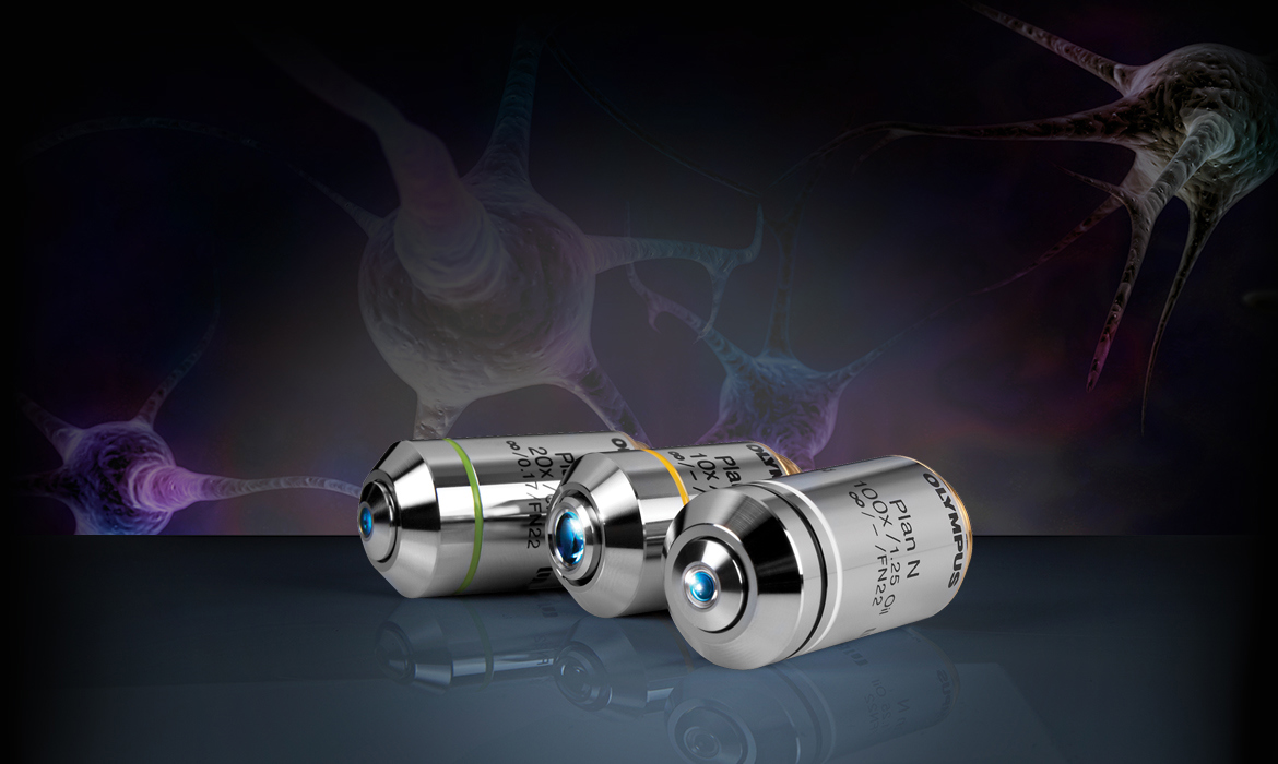
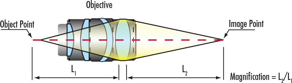
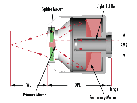
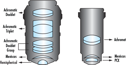
Multi-element objectives are critical for any microscopy technique, including many diagnostic techniques for the brain. Understanding the different types of objectives is important in order to ensure you are using the correct objective for your application.
Learn more about how to assemble a fluorescence microscopy system.
Below are common ailments of the brain that are detected by advanced diagnostic techniques such as fluorescence microscopy. Advancements in microscope objectives and other optical components allow these ailments to be more easily detected and treated.
Medical condition caused when blood supply to the brain is interrupted for an extended period of time, resulting in muscle weakness on one side of the body, loss of face control, numbness, and speech issues.
Progressive and incurable type of dementia that destroys memory and other important mental functions, beginning slowly and worsening over time.
Incurable disorder of the central nervous system (CNS) that affects movement and includes uncontrollable tremors.
Inherited, incurable condition that causes nerve cells in the brain to break down over time, causing jerky body movements and eventually the inability to talk.
Severe inflammation of the brain and spinal cord membranes that is typically onset by an infection and leads to fever, headache, and neck stiffness.
Condition characterized by recurring seizures mainly due to abnormal and increased electrical activity in the brain.
The most common type of traumatic brain injury, onset by heavy impact resulting in the brain shaking or moving inside the skull.
Cancerous or non-cancerous growths of abnormal cells in the brain with a variety of severity levels and types including astrocytoma, blastoma, ependymoma, and meningioma.


or view regional numbers
QUOTE TOOL
enter stock numbers to begin
Copyright 2024, Edmund Optics Singapore Pte. Ltd, 18 Woodlands Loop #04-00, Singapore 738100
California Consumer Privacy Acts (CCPA): Do Not Sell or Share My Personal Information
California Transparency in Supply Chains Act
This content may include material that has been generated or modified using artificial intelligence (AI).
The FUTURE Depends On Optics®